


Enhanced CAT image (University of Manchester)
Materials scientists at University of Manchester in the U.K. developed a faster and more feasible 3D color X-ray system with potential uses in health care, security inspections, and industrial quality assurance. The researchers, led by Manchester’s Robert Cernik, describe their invention in the current issue of the journal Analyst (free registration required).
Cernik’s team, which included colleagues from the Rutherford Appleton Laboratory also in the U.K., use standard X-ray computer tomography — sometimes called computed axial tomography or CAT scans — that take many X-ray views and combine them with an algorithm to provide a cross-sectional image of the subject. In health care, those images offer enhanced views of bones and soft tissue from various angles, but the technology is also applicable to industrial and security inspections.
The team developed a process for capturing all of the information in the X-ray beams called hyperspectral imaging, creating a more powerful X-ray method that before eluded practitioners and researchers alike. The new process makes possible hyperspectral imaging without the need for a synchrotron to produce the X-rays. The method notes Cernik “gives extra information about the material structure at each voxel — 3D equivalent of a pixel — of the 3D image. This extra information can be used to fingerprint the material present at each point in a 3D image.”
The new method also circumvents the need for silicon-based detectors. Cernik explains that silicon is a light atom that before could not stop high energy X-rays going through large objects. “Now we can achieve the same imaging results with an 80 by 80 pixel camera,” says Cernik, “made from cadmium zinc telluride, that supports real-time hyperspectral X-ray imaging up to very high energies.”
In addition, the new X-ray system produces 3D images faster than before. The researchers say the new system creates the composite image in one scanning motion taking several minutes, rather than the standard mapping process that collects and assembles many separate images.
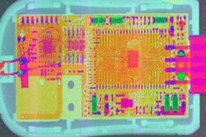
In the lab, the Manchester team used the system to examine a USB adapter that controls Web cams. They were able to identify the different elements and components inside the adapter by analyzing energy-sensitive radiographs and fluorescence patterns. The returned image (pictured at top) shows components highlighted in different colors to identify the elements bromine, barium, silver, tin, and zirconium.
Some of the first uses of the new X-ray system will likely be in health care. “The fact the image can be taken at the same time as using more conventional methods and on the same time scale means more information can be gathered from biopsy samples,” says Cernik. “This will more accurately differentiate between normal and abnormal tissue types reducing misdiagnosis.”
Other potential applications include security screening at airports to spot materials such as cocaine, semtex, precious metals, and radioactive substances. In addition, the researchers expect the technology can be used in aircraft maintenance, industrial inspections, and geophysical explorations. While the invention is still a lab-based system, Cernik is seeking industrial partners to further refine the X-ray technology for specific commercial applications.
Read more:
- 3-D, Low-Radiation Breast Cancer Imaging Technique Developed
- CT Image Analytics Adapted for COPD Diagnosis
- Low-Dose, High-Rez CT Scanning Technique Developed
- 4D Lung Imaging Technology Developed
- Faster Process Developed for Ultrasound Materials Testing
* * *
 RSS - Posts
You must be logged in to post a comment.