


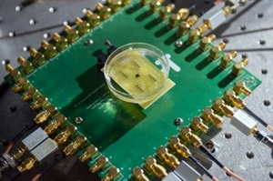
Heart imaging chip (Rob Felt, Georgia Institute of Technology)
19 February 2014. Engineers at Georgia Institute of Technology in Atlanta designed and lab-tested a microscopic chip to create real-time, three-dimensional images from inside the heart and blood vessels. The team led by mechanical engineering professor Levent Degertekin, with colleagues from Georgia Tech and Istanbul Technical University in Turkey, published its findings in this month’s issue of the journal IEEE Transactions on Ultrasonics, Ferroelectrics and Frequency Control (paid subscription required).
Degertekin and colleagues packed a system of ultrasound transducers — devices that convert signals from one form to another — on a single semiconductor chip 1.5 millimeters in diameter. The chip has 56 transmitting and 48 receiving elements arrayed around a tiny 430-micron hole for a guide wire. The chip also has power-saving circuits that allow the chip to operate with only 20 milliwatts of power, enough to energize the transducers, but also to reduce heat.
The Georgia Tech team designed the chip to be inserted into blood vessels and the heart through a catheter, and connect the chip to an external receiving device with 13 ultra-fine wires. This configuration would make the chip small and flexible enough to operate in the circulatory system, by incorporating some of the image processing on the chip itself, thus limiting the number of wires.
The researchers tested the chip inside the removed heart of a chicken under controlled conditions in their lab, and report transmitting 3-D images at 60 frames per second in real time, which they say is suitable quality and speed for catheter-based clinical applications. “This will give cardiologists the equivalent of a flashlight so they can see blockages ahead of them in occluded arteries,” says Degertekin in a university statement. “It has the potential for reducing the amount of surgery that must be done to clear these vessels.”
The next step for the team is to test the device in the lab with animals. If successful, they expect the university to license the technology to a medical device manufacturer for human clinical trials and FDA review.
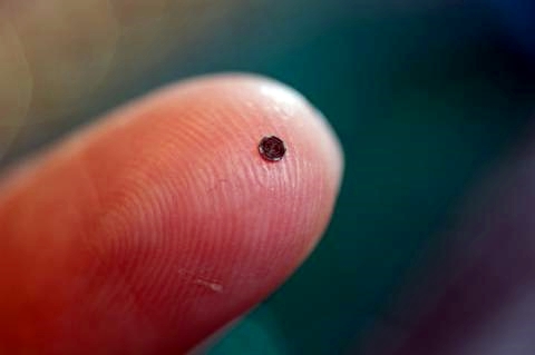
Heart imaging chip on fingertip (Rob Felt, Georgia Institute of Technology)
Read more:
- Heart Vessel Surgical Glue Shown Effective in Animal Tests
- Patch Finds More Heart Rhythm Problems Than Holter Monitor
- Biosensor Detects Brain-Damaging Proteins in Heart Surgery
- NIH Funding Micro-Sutures for Stem Cell Heart Muscle Repair
- Trial Testing External Power Connections for Heart Pumps
* * *
 RSS - Posts
You must be logged in to post a comment.