


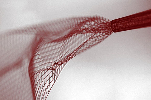
Microscopic image showing the soft wire mesh being injected through a glass needle less than 100 micrometers in diameter. (Lieber Research Group, Harvard University)
9 June 2015. Researchers at Harvard University developed a tiny electronic wire mesh that can be injected into the brain and demonstrated its diagnostic and therapeutic potential with lab mice. The team from the lab of chemistry professor Charles Lieber published its findings yesterday in the journal Nature Nanotechnology (paid subscription required, but full text available on the lab Web site). In addition, the university filed a patent on the invention and is seeking commercialization partners.
Lieber’s lab studies nanoscale materials, where 1 nanometer equals 1 billionth of a meter, including materials that interact with biological functions. In earlier work, the lab showed it was possible to grow nerve or cardiac tissue cells on embedded scaffolds, and record signals emitted by the cells. Lieber, with colleagues from Harvard and National Center for Nanoscience and Technology in Beijing, examined in this study if a nanoscale scaffolding could be inserted and interact with the brain at the cellular level.
The scaffold in this case is a tiny flexible wire mesh fitted between layers of polymer, fabricated with a process similar to etching microchips. Once formed on a surface, the covering layer dissolves, leaving the wire mesh that can be connected to external devices for sending or receiving signals. The soft wire mesh is then sucked into a microinjector, which compresses the mesh into a size that fits into a glass syringe needle, less than 100 micrometers in diameter.
The researchers first tested the injections of the mesh in simulations, using synthetic brain cells and hydrogels in the lab. When satisfied that the mesh would unfold from its compressed state in the syringe, the team injected the mesh into the lateral ventricle and hippocampus areas of the brains of live, anesthetized lab mice. A wire connecting to the mesh protruded from the skulls of the mice.
The findings show the wire mesh unfolded and was tolerated by the mice brain cells, with little immune rejection, and even integrates with the cells. High-resolution microscopy indicates the mice brain cells bind to and migrate along the injected mesh. In addition, the researchers can record brain activity from 16 electrodes on the mesh, which paper describes as well-defined signals.
“This opens up a completely new frontier where we can explore the interface between electronic structures and biology,” says Lieber in a university statement. “For the past thirty years, people have made incremental improvements in micro-fabrication techniques that have allowed us to make rigid probes smaller and smaller, but no one has addressed this issue — the electronics/cellular interface — at the level at which biology works.”
Read more:
- Patent Awarded for Neurostimulation Electronics
- Brain Sensor Designed for Wireless Connections
- Graphene Sensor Offers Clear Optical Access to Brain Cells
- University Starts Computer Science/Brain Research Consortium
- Consortium to Develop Common Neuroscience Data Format
* * *
 RSS - Posts
You must be logged in to post a comment.