


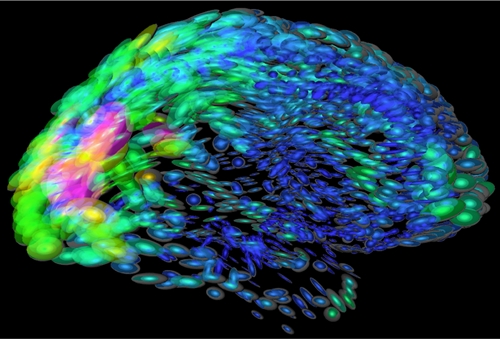
(Arthur Toga, UCLA/NIH.gov)
17 December 2015. An engineering group at University of Arizona is developing a new technology that promises to provide better images of electrical activity in the brain. The project, led by Arizona biomedical engineering professor Russell Witte, is funded by a three-year, $1.15 million grant from National Institute of Neurological Disorders and Stroke, part of National Institutes of Health.
Witte and colleagues seek to provide more detailed, real-time images that record electrical impulses among neurons or nerve cells in the brain. The undertaking is a multi-disciplinary effort, with contributions from specialists in psychology, neurosurgery, neurology, emergency medicine, and mathematics at Arizona and three other institutions. The technology is expected to improve diagnosis and treatment of neurological disorders such as epilepsy, Parkinson’s disease, and traumatic brain injury.
“We know very little about how neurons act collectively to guide our thoughts, emotions and behaviors, or cause seizures or mood swings,” says Witte in a university statement.” Functional magnetic resonance imaging, or fMRI, and electroencephalography, or EEG offer clues, notes Witte, but produce images with poor resolution. “We think our new technology could overcome that limitation.”
That new technology adapts ultrasound emitting high-frequency sound waves that interact with tissue and organs, and return images of activity inside the body in real time. The university says Witte’s lab already developed an ultrasound technique to better diagnose irregular heart rhythms, and is starting up a new company to commercialize that technique.
In this new project, the team plans to take ultrasound further, to interact with electrical currents in the brain as well as tissue. This noninvasive technology known as acoustoelectric brain imaging is expected to mix ultrasound with neural impulses to produce unique voltage modulations acting as electronic signatures of brain activity. Electrodes placed on the skull would then record those signatures.
With this technology, say the researchers, clinicians could send an acoustoelectric signal through the brain that returns an accurate real-time map of brain functions. To achieve this goal, however, several obstacles need to be overcome, including detecting weak acoustoelectric signals through the skull while still maintaining the patient’s safety. The team plans to build a lab model for the technology’s design and initial testing, as well as further tests with rat and pig brains.
If successful, the technology would enable clinicians to observe brain activity that can lead to more precise removal of brain tissue causing seizures in people with epilepsy. The technology could also be used to monitor the effects of drugs to reduce hallucinations, indicated by spikes in electrical activity at various points in the brain. Current imaging technologies record images every two seconds, but at this early stage, the researchers still do not know the speed or extent of images produced by acoustoelectric brain imaging.
This project is funded under the U.S. inter-agency BRAIN Initiative — short for Brain Research through Advancing Innovative Neurotechnologies — that seeks new technologies to “produce real-time pictures of complex neural circuits and visualize the rapid-fire interactions of cells that occur at the speed of thought.”
Read more:
- Advocacy Group, Genetics Company Partner on Brain Disorder
- Brain Tissue Banks Partner on Autism Research
- Delivery Technique Devised to Cross Blood-Brain Barrier
- Spin-Off Formed to Develop Brain Disorder Therapies
- Wireless System Developed to Deliver Drugs to Brain
* * *
 RSS - Posts
You must be logged in to post a comment.