


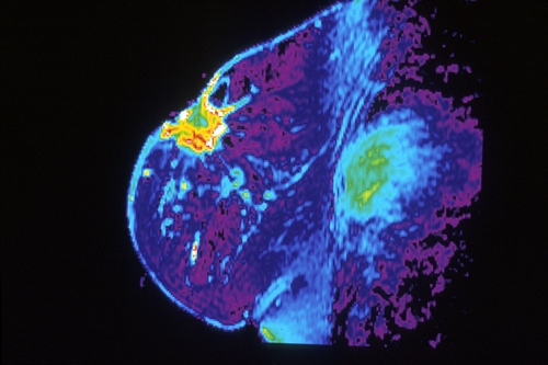
MRI of breast (National Cancer Institute)
27 September 2017. A large-scale clinical trial is underway comparing the effectiveness of two-dimensional and three-dimensional mammograms in screening for breast cancer. The Tomosynthesis Mammographic Imaging Screening Trial or Tmist is enrolling 165,000 healthy women to determine if the more technologically advanced 3-D mammograms reduce the chance for women to develop advanced, life-threatening cancer than 2-D mammogram images.
Tomosynthesis mammographic imaging is a digital technique that constructs a composite 3-D image of the breast from multiple X-rays taken at different angles. The images provide a fuller picture of the breast for detecting denser tissue indicating possible cancer than conventional 2-D images taken from two angles. Both imaging techniques are approved by the Food and Drug Administration, but the trial seeks to find out if the more advanced technology is also more effective in detecting potentially dangerous breast cancer cases.
The trial is conducted by Ecog-Acrin Cancer Research Group, an organization that designs and undertakes biomarker-driven cancer research with adults who have or are at risk of developing cancer. The group formed from a merger of Eastern Cooperative Oncology Group (Ecog) and the American College of Radiology Imaging Network (Acrin), and is part of National Clinical Trials Network. National Cancer Institute, part of National Institutes of Health, is a partner in and funder of the study.
“We need to determine if 3-D mammography is better than 2-D at finding the sort of breast cancers that are most likely to spread and kill women,” says study chair Etta Pisano in an Ecog-Acrin statement. “If a newer screening technology does not reduce the numbers of advanced, life-threatening cancers, then are we really improving screening for breast cancer?” Pisano is a radiologist at Beth Israel Deaconess Medical Center and professor of radiology at Harvard Medical School in Boston.
Tmist that opened in July is recruiting 165,000 healthy women, age 45 to 74, to take part at 100 locations in the U.S. Participants, who planned to to have routine mammograms, will be randomly assigned to receive 2-D or 3-D images for 5 years. Most participants will have mammograms taken every year, although some women in lower-risk groups for developing breast cancer will have their mammograms every other year. Women taking part in the trial will then be tracked for a total of 8 years by the research team, through reviews of participants’ medical records and in some cases with follow-up phone surveys.
In addition, the researchers are asking participants to provide voluntary blood samples and mouth swabs to build a bio-repository for future studies of genetic markers associated with breast cancer. Tissue samples from biopsies, if taken, will also be collected.
The clinical trial is looking primarily at the percentage of participants diagnosed with advanced cases of breast cancer during the full study period. The study is also assessing several related factors in breast cancer diagnosis, including evaluation of breast images, agreement between local and centralized pathology reviews of biopsies, and overall health care costs covering both diagnostics and cancer care over the study period.
Ecog-Acrin says the 100 mammography sites in Tmist are geographically distributed to attract a diverse ethnic and racial mix in participants. Several Canadian mammography clinics enrolled some 3,000 participants in a similar study conducted earlier to help design Tmist.
More from Science & Enterprise:
- App Screens for Liver Diseases with Eye Selfies
- FDA Clears Algorithm-Assisted Cancer Diagnosis
- Lab-On-Chip Quickly Isolates, Captures Exosomes
- Liquid Biopsy Company Gains $360M for Cancer Sequencing
- Crispr-Based Diagnostics System Designed
* * *
 RSS - Posts
You must be logged in to post a comment.