


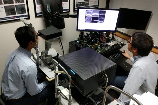
High-resolution retinal scanner (Simon Fraser University)
22 December 2017. A Canadian engineering lab designed a retinal scanner that helps diagnose eye diseases in their early stages, often well before vision loss occurs. The scanner is an invention of Marinko Sarunic, a professor of biomedical engineering at Simon Fraser University in Burnaby, British Columbia.
Sarunic’s lab studies biomedical optics, specializing in optical coherence tomography, or OCT, a diagnostic technique that scans tissue in the eye. Current OCT technologies use low-resolution images to identify already dead or highly damaged tissue in the retina, a layer on the back of the eye with light-sensitive cells that convert light into neural signals, carried into the brain by the optic nerve.
Retinal damage is among the leading causes of vision loss in North America. Centers for Disease Control and Prevention says some 21 million people in the U.S. have vision problems from disorders such as age-related macular degeneration or AMD, diabetic retinopathy, and glaucoma, leading to blindness in 3.4 million individuals. Another 1 million Canadians suffer from vision loss due to these eye diseases, according to the university.
In the new system, the Simon Fraser team is developing a high-resolution device that produces 3-D images of the retina, detecting early signs of damage to the light-sensitive cells and the small blood vessels supporting the cells. The system fits on a desktop, which should help make it practical for smaller hospitals and clinics.
Sarunic and colleagues describe the scanner in the November 2017 issue of the Journal of Biomedical Optics (paid subscription required). The system is non-invasive, using near-infrared light scans to achieve micrometer resolution, providing both large and small fields of view. The smaller field of view makes it possible to return detailed images of photoreceptors — the light-sensitive cells in the retina — as well as fine blood vessels in the retina. The article also reports on tests with 4 healthy volunteers, demonstrating its functions and utility in the clinic.
“It’s a breakthrough in clinical diagnostics,” says Sarunic in a university statement. “With the high-resolution scanner, ophthalmologists and optometrists can detect damage and changes to small numbers of individual photoreceptors, giving them a diagnosis before the patient loses vision, and the potential to take preventative measures.”
Vancouver ophthalmologist and retinal specialist Eduardo Navajas adds that the scanner simplifies early detection, removing the current need for dye injections into the eye. “Dr. Sarunic’s new imaging technology,” says Navajas, “is benefiting patients, allowing us to diagnose and treat wet AMD and diabetic eye disease before patients develop bleeding and permanent damage to their retina.”
More from Science & Enterprise:
- Contact Lens Devised to Measure Glaucoma Eye Pressure
- Preclinical Data Show Promise for Diabetic Eye Drug
- Synthetic Retina Developed with Natural Materials
- Trial to Test Gene Therapy for Inherited Eye Disease
- Trial Testing Retina Cell Implants for Glaucoma
* * *
 RSS - Posts
You must be logged in to post a comment.