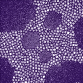
Transmission electron microscopy image of nanoparticles designed for deep-tissue imaging. (Zhipeng Li, University at Buffalo)
Researchers in the U.S., Sweden, China, and Korea created illuminated nanoscale particles that can be detected through a 3.2 centimeter, or 1.26 inch layer of tissue. The team led by University at Buffalo, New York chemistry professor Paras Prasad and University of Massachusetts medical professor Gang Han published its findings last month in the journal ACS Nano (paid subscription required).
Prasad, Han and colleagues say their discovery can meet the need for a high-resolution, high-contrast optical bioimaging technology to explore deeper in human tissue, such as to identify cancerous tumors. The technology uses nanoscale particles — one nanometer equals one billionth of a meter — that absorb and emit near-infrared light, with the emitted light having a much shorter wave length than the absorbed light. This property makes it possible to get deeper and higher-contrast images than current fluorescence-based methods.
The particles first absorb pairs of photons, then combine them into single, higher-energy photons that are then emitted. This absorption and emission process is done in the near infrared (NIR) region of the electromagnetic spectrum. This part of the spectrum helps reduce background interference, and for biological tissue, absorbs and scatters the least light in this range.
The team’s nanoparticles are made of a crystalline core with thulium, sodium, ytterbium, and fluorine, encased inside a square, calcium-fluoride shell. Calcium fluoride is found in bone and tooth mineral, making the particles bio-compatible, and thus reducing the risk of adverse effects. The researchers also found the shell helps increase the illumination efficiency of the encased particles.
The team tested the nanoparticles by first injecting them in mice, then placed in a capsule behind a 3.2 centimeter layer of pork. In both cases, the researchers were able to obtain high-contrast images of the particles shining through tissue.
The next step for the researchers is to explore ways of finding and attaching the nanoparticles to cancer cells and other biological targets that could be imaged.
Read more:
- Buffalo, Zimbabwe Universities Partner on Nanotech Medicines
- Polymer Nanoparticles Tested to Respond,Treat Inflammation
- University Consortium to Research Nanotech Health Monitors
- RNA Nanoparticles Advanced for Cancer Drug Delivery
- University to Develop, Commercialize HIV/AIDS Nanomedicines
* * *

 RSS - Posts
You must be logged in to post a comment.