


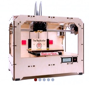
Replicator 3-D printer (MakerBot Industries LLC)
Researchers at University of Notre Dame in Indiana developed a process for three-dimensional printing of anatomical models from computed tomography (CT) scans. The team from the lab of Matthew Leevy, a research professor at Notre Dame’s Integrated Imaging Facility, published its findings online last week in text and video form in the Journal of Visualized Experiments.
The idea for the project came from Evan Doney, an undergraduate engineering student and first author of the paper, who was joined by several colleagues at Notre Dame, as well as a representative of MakerBot Industries LLC, a 3-D printing company in Brooklyn, New York. Doney and colleagues reported on scans and printing of skeletal and organ models of anesthesized lab rats and a preserved skull of a New Zealand white rabbit.
CT scans — sometimes called CAT scans — use X-rays to visualize bones, soft tissue, and organs. The scans take a rotating image of successive thin slices of the body that are then reconstructed with software and displayed on a computer monitor. The Notre Dame researchers used an Albira CT scanner made by Carestream Molecular Imaging in Connecticut.
The Albira Reconstructor software, say the researchers, was able to return images and associated data with sufficient resolution for the 3-D printers to produce at least some models. The team used three different printers to demonstrate the process’s interoperability with different hardware platforms: MakerBot, Shapeways, and ProJet HD 3000.
The paper notes that the Albira software provided enough data for the printers to create skeletal features of the scanned animals, but not the organ tissue, which required considerably more processing. The data needed conversion to other software formats for further analysis and to provide enough detail for surface rendering. Two other programs were needed to smooth and repair mesh models.
Leevy notes that the university is exploring markets for 3-D models made from imaging data, including being able to make models from data not identified from visual inspection. “Not only can we print bone structure,” says Leevy, “but we’re starting to collect patient data and print out the anatomical structure of patients with different disease states to aid doctors in surgical preparation.”
Read more:
- Color X-Ray System Devised for Health, Security, Industry
- Dual-Purpose Medical Imaging Contrast Agent Developed
- 3-D, Low-Radiation Breast Cancer Imaging Technique Developed
- CT Image Analytics Adapted for COPD Diagnosis
- Purdue, Adobe Create Process to Strengthen 3-D Print Objects
* * *
 RSS - Posts
[…] Anatomical Models 3-D Printed from Tomography Scans […]