


Electrosurgery (Imperial College London)
Medical technology researchers from Hungary and the U.K. developed a device that analyzes the smoke-like aerosol released during cancer electrosurgery to determine if the dissected tissue is cancerous. The team from Imperial College London led by medical faculty member Zoltán Takáts published its findings in today’s issue of the journal Science Translational Medicine (paid subscription required).
Takáts and colleagues from Imperial College London, University of Debrecen in Hungary, and the company MediMass Ltd. in Budapest designed a tool for use during solid tumor surgery — e.g., breast or lung tumors — where samples of tissues are visually inspected by surgeons or extracted for testing by pathologists. If the sample needs to be tested, under current procedures the patient remains under general anesthesia while a separate lab analyzes the tissue, a process that can take as long as a half hour. The uncertainty of current methods can result in the need for a second operation to fully remove the cancerous tissue, which the university says happens to one in five breast cancer patients who have surgery.
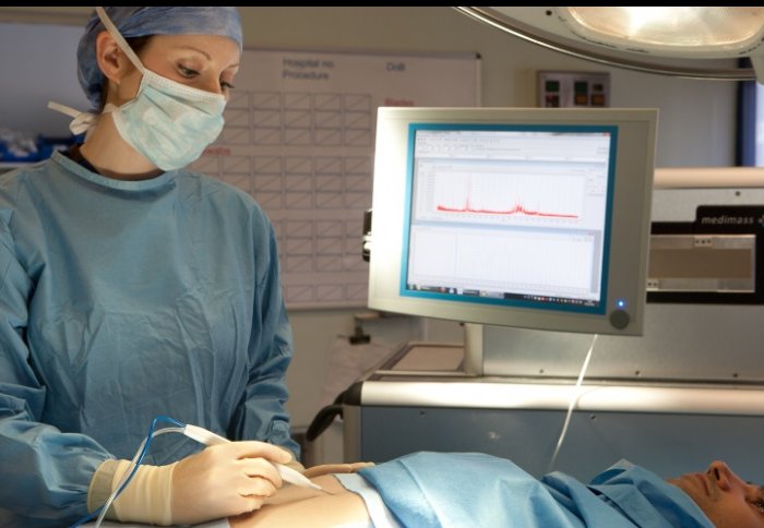
The researchers created an “intelligent knife,” or iKnife, for use in electrosurgery, an established technology that applies a concentrated electric current to quickly heat diseased tissue, which makes it possible to cut the tissue while minimizing the loss of blood. During this technique, the heated tissue releases a smoke-like aerosol plume that provides the raw material analyzed by the iKnife.
To analyze the plume, the research team attached sensors from a mass spectrometer to the electrosurgical scalpel to detect the chemical composition of the plume. The readings from the iKnife are then analyzed by the attached mass spectrometer to determine the nature of the cells being heated during electrosurgery.
Takáts and colleagues first analyzed tissue samples with the iKnife from 302 brain, lung, breast, stomach, colon, and liver cancer patients, generating a reference library of 1,624 cancerous and 1,309 noncancerous entries. The team then used the iKnife to collect real-time samples from 81 cancer patients, and identifications made from the samples taken during surgery all matched the post-surgical evaluations using current methods. The real-time diagnoses took 2 to 3 seconds, compared to the 20 to 30 minutes currently needed.
One drawback to the iKnife, however, is the need for people other than the surgeons to read the mass spectrometer to determine the nature of the tissue. The researchers hope to devise a test of the technology to determine if providing surgeons with real-time access to the analytical data improves patient outcomes.
The paper’s first author, Júlia Balog, is a neurobiologist and computer scientist with MediMass Ltd., a company in Budapest founded in 2008 to commercialize the iKnife technology. Takáts serves as the company’s chief scientist.
Read more:
- Challenge Seeks Point-of-Care Staph Bacteria Detector
- Device Detects Residual Cancer in Lumpectomy Tissue
- First Bioengineered Vein Implanted for U.S. Dialysis Patient
- Surgical Agents Developed for Biopsies in Confined Spaces
- Technique Calculates X-Rays for Minimally Invasive Surgery
* * *
 RSS - Posts
You must be logged in to post a comment.