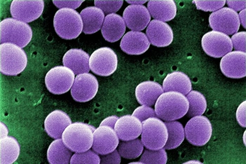
10 June 2016. A biomedical engineering center at Harvard University developed a process for quickly isolating staph bacteria from clinical samples for lab testing. The team led by Donald Ingber, director of the Wyss Institute for Biologically Inspired Engineering at Harvard, published its findings earlier this week in the journal PLoS One.
Ingber and colleagues developed the new technique to make it easier to discover Staphylococcus aureus, or staph bacteria, and other pathogens causing infections contracted in hospitals and clinics. Staph bacteria are associated with osteoarthritis infections, sometimes resulting from joint replacements, where infections can lead to painful inflammation. Samples of tissue or joint fluid from patients, however, are complex and pathogens in theses samples are often difficult to isolate and identify.
The Wyss team, with former institute colleagues in France, adapted a process designed earlier to clean blood of sepsis bacteria, causing dangerous infections. That method uses an engineered molecule called FcMBL acting like a natural protein known as mannose-binding lectin that binds to carbohydrates found in a broad range of bacteria and viruses including those associated with sepsis. Wyss researcher Michael Super, a co-author of the new study, genetically engineered mannose-binding lectin to combine with molecules from the Fc region in antibodies that extends their persistence in blood serum.
As reported in Science & Enterprise, Ingber and Super founded Opsonix Inc., a spin-off company to commercialize the sepsis-cleaning technology. That technology includes coating nanoscale beads with the engineered mannose-binding lectin, then magnetizing the coated beads to attract microbes and toxins.
When the researchers tried those techniques to isolate staph bacteria in tissue samples from a biobank of osteoarthritis patients, they discovered the magnetized and coated beads cannot bind directly to bacteria in the joint fluids. One reason is the viscosity of the joint fluid, but also the presence of immune cells and proteins in the samples that mask carbohydrate molecules on bacteria that serve as binding targets for FcMBL.
To improve the performance and efficiency of the beads, the researchers wash the clinical samples in an enzyme cocktail before exposure to the FcMBL. The pre-treatment cocktail is made of proteases, enzymes that break down the peptides holding together the amino acids in proteins masking the binding carbohydrates. The pre-treatment also uses hyaluronidase, another enzyme that helps break down the viscous nature of the joint fluids.
The combination of pre-treatment and FcMBL beads makes it possible to isolate the staph bacteria from the samples within 2 hours. Once isolated, the bacteria or other pathogens can then be tested with standard lab procedures.
Ingber says in a Wyss Institute statement that the technique can be extended to other pathogens and specimen samples, such as blood, urine, sputum, and cerebral spinal fluid. “In addition to saving more lives,” adds Ingber, “this new method also should reduce the use of broad-spectrum antibiotic therapies, or sub-optimal regimens, and thereby decrease development of antibiotic-resistant organisms that become a more general threat in the long run.”
Read more:
- More Genomic Tests Enhance Personalized Cancer Care
- Venture Firm Funding Univ. Blood Diagnostics Technology
- Start-Up Licenses Genetics Technology for HIV Diagnostics
- Genomic Variations Reveal Cholesterol, Heart Disease Risks
- NIH Funds Biosensors to Monitor Oxygen in Tissue
* * *


 RSS - Posts
[…] Process Devised to Quickly Isolate Bacteria in Lab Samples […]