


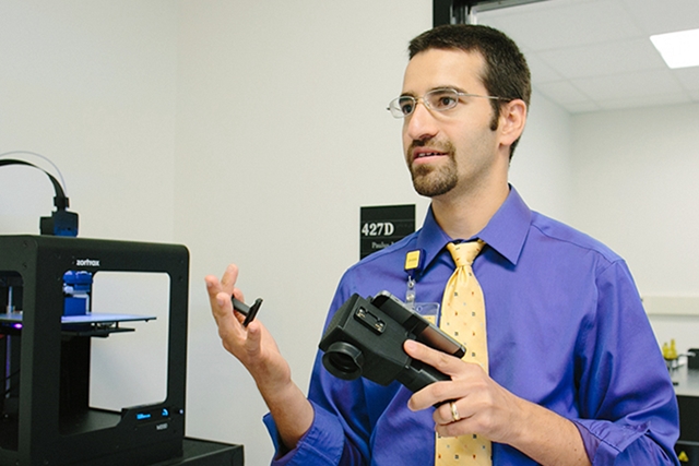
Yannis Paulus holds a RetinaScope device (University of Michigan)
30 Apr. 2019. An ophthalmology lab at University of Michigan developed a smartphone-based system that speeds screening for an eye disease resulting from diabetes. A team led by Michigan medical school professor Yannis Paulus discussed its RetinaScope system at the Association for Research in Vision and Ophthalmology annual meeting, now underway in Vancouver, British Columbia.
High levels of blood sugar can cause serious complications in people with diabetes, including vision problems. Among these issues is damage to the fine blood vessels in the retina, located in the back of the eye that detects and converts light to signals sent through the optic nerve to the brain. Diabetic retinopathy is the name given to this condition that can range from mild leakage in the eye to swelling and distortion of the blood vessels, and proliferation of new blood vessels to compensate for the damage.
The disease can be detected by trained clinicians using a retinal camera, but the equipment is expensive and requires specialized installation, with turnaround time for interpreted results taking from 2 to 7 days. In addition, retinal cameras and their trained operators are found in relatively few specialized eye clinics. The Michigan team, with colleagues from Washington University in St. Louis and University of California in Berkeley, are seeking a technology that primary care physicians can use to screen for diabetic retinopathy, or D.R., then refer patients to specialists if needed.
“The key to preventing D.R.-related vision loss is early detection through regular screening,” says Paulus in a university statement. “We think the key to that is bringing portable, easy-to-administer, reliable retinal screening to primary care doctors’ offices and health clinics.”
The RetinaScope system attaches a compact, high-resolution camera with wide-angle images like those returned by retinal cameras to a smartphone. The combination of camera and phone make it possible for a hand-held device to capture images comparable to the large, bulky conventional retinal cameras.
The next challenge is to speed-up the analysis of captured retinal images. For that task, the RetinaScope system uses software called EyeArt developed by Eyenuk Inc. in Woodland Hills, California. EyeArt software employs deep neural networking, a form of artificial intelligence, or A.I., trained with some 2 million retinal images worldwide from 500,000 patients. Images from RetinaScope are uploaded to Eyenuk databases in the cloud, where EyeArt analyzes the images and returns results to the smartphone in about 1 minute.
“Deep neural network is an A.I. software platform,” notes Paulus, “that can enhance and review images and provide automated grading of lesions present in D.R., indicating which lesions require referral to an ophthalmologist for follow-up.”
Paulus and colleagues recruited 69 patients from the university’s eye clinic to evaluate the RetinaScope system, with help from 2 trained clinicians reviewing the same images. After participants’ pupils were dilated — increased in size for better examination of the eye and optic nerve — they were screened for diabetic retinopathy by the RetinaScope system, with results returned indicating a recommendation for follow-up by a specialist or no referral warranted. The participants earlier received conventional slit-lamp eye exams.
The results show the RetinaScope system correctly detected indicators of diabetic retinopathy 87 percent of the time, higher than the 80 percent threshold recommended for eye screening devices, with the ability to accurately detect the absence of disease indicators 73 percent of the time. One of the clinicians also evaluating the images correctly detected diabetic retinopathy indicators 96 percent of the time, more than the RetinaScope system, but both clinicians accurately identified the absence of the disease no more than 47 percent of the time.
“This is the first time A.I. used on a smartphone-based platform has been shown to be effective when compared to the gold standard of clinical evaluation,” says Paulus. The RetinaScope team now plans to enhance the system to enable its use without pupil dilation, and to advance the technology to FDA for review.
More from Science & Enterprise:
- Trials OK’d for Inherited Eye Disease Therapy with Crispr
- Trials to Test Stem Cell Treatments for Eye Injuries
- Hand-Held Device Inspects Individual Retina Cells
- Allergan Licensing Crispr for Inherited Eye Disease
- FDA Clears A.I. Software Detecting Diabetic Eye Disease
* * *
 RSS - Posts
[…] Smartphone System Detects Diabetic Eye Disease […]