


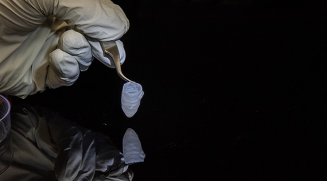
Left-ventricle heart model ((Luke MacQueen and Michael Rosnach, Harvard University)
24 July 2018. A university bioengineering group developed a model of a left ventricle, the heart chamber that pumps blood to the body, made with engineered heart muscle tissue to replace animals for drug testing. A team from the lab of Harvard University biophysics and engineering professor Kevin Kit Parker describe the device in yesterday’s issue of the journal Nature Biomedical Engineering (paid subscription required).
Parker’s team, with colleagues from other Harvard labs and affiliated hospitals in Boston, are seeking more reliable methods of testing new treatments for heart disease that up to now rely on animal testing for drug toxicity and effectiveness before testing with humans. Heart disease is a widespread problem in the U.S. According to Centers for Disease Control and Prevention, some 610,000 people each year die of heart disease, accounting for 1 in every 4 deaths, the leading cause of death for both men and women. A common form of heart disease is atrial fibrillation, the leading type of heart arrhythmia, where the heart rhythm becomes irregular and blood does not pump evenly from the upper chambers, or atria, to the lower ventricle chambers of the heart. From 3 to 6 million Americans have atrial fibrillation.
The researchers devised a scale model, about 1/250th the size of a human heart. The device is built around a scaffold of nanoscale fibers arrayed in parallel, similar to the human heart. These materials are fabricated from tiny droplets of liquid biodegradable polymer and gelatin compounds, fed through a jet needle, where they solidify and are spun into fibers. The fibers are then captured around a bullet-shaped collector, which aligns the fibers in the scaffold similarly to a human heart.
“It is important to recapitulate the structure of the natural muscle to obtain ventricles that function like their natural counterparts,” says Luke MacQueen, postdoctoral researcher and first author of the paper in a university statement. “When the fibers are aligned, the cells will be aligned, which means they will conduct and contract the way that native cells do.”
The team seeded the scaffold with stem and precursor cells of rat and human heart muscles, which started forming tissue and began beating in a synchronous rhythm in 3 to 5 days. As tissue began building up around the scaffold, researchers could then insert a catheter to measure calcium propagation through the model — a key indicator of heart muscle health — and pressure. In this form, the team could perform immediate tests of the device in a petri dish, including simulation of a heart attack.
For longer-term effects, the researchers needed a bioreactor device to sustain the left ventricle model for extended periods of time. The bioreactor has separate sections with inserts for valves and catheters, which supports heart models made with tissue derived from human stem cells for as long as 6 months. The team tested a simulated case of arrhythmia in the bioreactor model to prove the concept. “The fact that we can study this ventricle over long periods of time is really good news for studying the progression of diseases in patients as well as drug therapies that take a while to act,” notes MacQueen.
The researchers next plan to test the ventricle model with stem cells derived from patients with heart disease. Harvard University filed for patent protection on the device and is seeking commercialization partners.
Ventricle heart model beating (Luke MacQueen, Harvard University)
More from Science & Enterprise:
- Computer-Aided Bone Graft Process Devised with Stem Cells
- Smart Bandage Monitors, Provides Drugs to Chronic Wounds
- University Tissue, Cell Fabrication Lab Set to Open
- Electrodes for Implants 3-D Printed on Soft Materials
- Company Claims High Speeds, Resolution in 3-D Printed Tissue
* * *
 RSS - Posts
[…] Tissue-Engineered Heart Chamber Model Developed […]